| dc.contributor.author | Le Gros, Mark A. | |
| dc.contributor.author | Clowney, E. Josephine | |
| dc.contributor.author | Magklara, Angeliki | |
| dc.contributor.author | Yen, Angela | |
| dc.contributor.author | Markenscoff-Papadimitriou, Eirene | |
| dc.contributor.author | Colquitt, Bradley | |
| dc.contributor.author | Myllys, Markko | |
| dc.contributor.author | Kellis, Manolis | |
| dc.contributor.author | Lomvardas, Stavros | |
| dc.contributor.author | Larabell, Carolyn A. | |
| dc.date.accessioned | 2017-04-03T16:35:52Z | |
| dc.date.available | 2017-04-03T16:35:52Z | |
| dc.date.issued | 2016-11 | |
| dc.date.submitted | 2016-08 | |
| dc.identifier.issn | 22111247 | |
| dc.identifier.uri | http://hdl.handle.net/1721.1/107824 | |
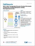
| dc.description.abstract | Summary - The realization that nuclear distribution of DNA, RNA, and proteins differs between cell types and developmental stages suggests that nuclear organization serves regulatory functions. Understanding the logic of nuclear architecture and how it contributes to differentiation and cell fate commitment remains challenging. Here, we use soft X-ray tomography (SXT) to image chromatin organization, distribution, and biophysical properties during neurogenesis in vivo. Our analyses reveal that chromatin with similar biophysical properties forms an elaborate connected network throughout the entire nucleus. Although this interconnectivity is present in every developmental stage, differentiation proceeds with concomitant increase in chromatin compaction and re-distribution of condensed chromatin toward the nuclear core. HP1β, but not nucleosome spacing or phasing, regulates chromatin rearrangements because it governs both the compaction of chromatin and its interactions with the nuclear envelope. Our experiments introduce SXT as a powerful imaging technology for nuclear architecture. | en_US |
| dc.description.sponsorship | National Institutes of Health (U.S.) (grant R01DA030320) | en_US |
| dc.description.sponsorship | National Institutes of Health (U.S.) (grant U01DA040582) | en_US |
| dc.description.sponsorship | National Center for X-ray Tomography (Lawrence Berkeley National Laboratory) ( NIH (P41GM103445) | en_US |
| dc.description.sponsorship | United States. Department of Energy. Office of Biological and Environmental Research (DE-AC02-5CH11231) | en_US |
| dc.language.iso | en_US | |
| dc.publisher | Elsevier | en_US |
| dc.relation.isversionof | http://dx.doi.org/10.1016/j.celrep.2016.10.060 | en_US |
| dc.rights | Creative Commons Attribution-NonCommercial-NoDerivs License | en_US |
| dc.rights.uri | http://creativecommons.org/licenses/by-nc-nd/4.0/ | en_US |
| dc.source | Elsevier | en_US |
| dc.title | Soft X-Ray Tomography Reveals Gradual Chromatin Compaction and Reorganization during Neurogenesis In Vivo | en_US |
| dc.type | Article | en_US |
| dc.identifier.citation | Le Gros, Mark A., E. Josephine Clowney, Angeliki Magklara, Angela Yen, Eirene Markenscoff-Papadimitriou, Bradley Colquitt, Markko Myllys, Manolis Kellis, Stavros Lomvardas, and Carolyn A. Larabell. “Soft X-Ray Tomography Reveals Gradual Chromatin Compaction and Reorganization During Neurogenesis In Vivo.” Cell Reports 17, no. 8 (November 2016): 2125–2136. | en_US |
| dc.contributor.department | Massachusetts Institute of Technology. Computer Science and Artificial Intelligence Laboratory | en_US |
| dc.contributor.department | Massachusetts Institute of Technology. Department of Electrical Engineering and Computer Science | en_US |
| dc.contributor.mitauthor | Yen, Angela | |
| dc.contributor.mitauthor | Kellis, Manolis | |
| dc.relation.journal | Cell Reports | en_US |
| dc.eprint.version | Final published version | en_US |
| dc.type.uri | http://purl.org/eprint/type/JournalArticle | en_US |
| eprint.status | http://purl.org/eprint/status/PeerReviewed | en_US |
| dspace.orderedauthors | Le Gros, Mark A.; Clowney, E. Josephine; Magklara, Angeliki; Yen, Angela; Markenscoff-Papadimitriou, Eirene; Colquitt, Bradley; Myllys, Markko; Kellis, Manolis; Lomvardas, Stavros; Larabell, Carolyn A. | en_US |
| dspace.embargo.terms | N | en_US |
| dc.identifier.orcid | https://orcid.org/0000-0001-5951-9358 | |
| mit.license | PUBLISHER_CC | en_US |
