| dc.contributor.author | Brielle, Shlomi | |
| dc.contributor.author | Kaganovich, Daniel | |
| dc.contributor.author | Gura Sadovsky, Rotem | |
| dc.contributor.author | England, Jeremy L. | |
| dc.date.accessioned | 2017-06-13T19:27:42Z | |
| dc.date.available | 2017-06-13T19:27:42Z | |
| dc.date.issued | 2017-03 | |
| dc.date.submitted | 2016-12 | |
| dc.identifier.issn | 2211-1247 | |
| dc.identifier.uri | http://hdl.handle.net/1721.1/109838 | |
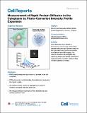
| dc.description.abstract | The fluorescence microscopy methods presently used to characterize protein motion in cells infer protein motion from indirect observables, rather than measuring protein motion directly. Operationalizing these methods requires expertise that can constitute a barrier to their broad utilization. Here, we have developed PIPE (photo-converted intensity profile expansion) to directly measure the motion of tagged proteins and quantify it using an effective diffusion coefficient. PIPE works by pulsing photo-convertible fluorescent proteins, generating a peaked fluorescence signal at the pulsed region, and analyzing the spatial expansion of the signal. We demonstrate PIPE’s success in measuring accurate diffusion coefficients in silico and in vitro and compare effective diffusion coefficients of native cellular proteins and free fluorophores in vivo. We apply PIPE to measure diffusion anomality in the cell and use it to distinguish free fluorophores from native cellular proteins. PIPE’s direct measurement and ease of use make it appealing for cell biologists. | en_US |
| dc.language.iso | en_US | |
| dc.publisher | Elsevier | en_US |
| dc.relation.isversionof | http://dx.doi.org/10.1016/j.celrep.2017.02.063 | en_US |
| dc.rights | Creative Commons Attribution-NonCommercial-NoDerivs License | en_US |
| dc.rights.uri | http://creativecommons.org/licenses/by-nc-nd/4.0/ | en_US |
| dc.source | Elsevier | en_US |
| dc.title | Measurement of Rapid Protein Diffusion in the Cytoplasm by Photo-Converted Intensity Profile Expansion | en_US |
| dc.type | Article | en_US |
| dc.identifier.citation | Gura Sadovsky, Rotem; Brielle, Shlomi; Kaganovich, Daniel; England and Jeremy L. “Measurement of Rapid Protein Diffusion in the Cytoplasm by Photo-Converted Intensity Profile Expansion.” Cell Reports 18, no. 11 (March 2017): 2795–2806 © 2017 The Authors | en_US |
| dc.contributor.department | Massachusetts Institute of Technology. Computational and Systems Biology Program | en_US |
| dc.contributor.mitauthor | Gura Sadovsky, Rotem | |
| dc.contributor.mitauthor | England, Jeremy L. | |
| dc.relation.journal | Cell Reports | en_US |
| dc.eprint.version | Final published version | en_US |
| dc.type.uri | http://purl.org/eprint/type/JournalArticle | en_US |
| eprint.status | http://purl.org/eprint/status/PeerReviewed | en_US |
| dspace.orderedauthors | Gura Sadovsky, Rotem; Brielle, Shlomi; Kaganovich, Daniel; England, Jeremy L. | en_US |
| dspace.embargo.terms | N | en_US |
| dc.identifier.orcid | https://orcid.org/0000-0001-9863-8616 | |
| dc.identifier.orcid | https://orcid.org/0000-0001-8414-3153 | |
| mit.license | PUBLISHER_CC | en_US |