| dc.contributor.author | Caron, Tyler J | |
| dc.contributor.author | Scott, Kathleen E | |
| dc.contributor.author | Sinha, Nishita | |
| dc.contributor.author | Muthupalani, Sureshkumar | |
| dc.contributor.author | Baqai, Mahnoor | |
| dc.contributor.author | Ang, Lay-Hong | |
| dc.contributor.author | Li, Yue | |
| dc.contributor.author | Turner, Jerrold R | |
| dc.contributor.author | Fox, James G | |
| dc.contributor.author | Hagen, Susan J | |
| dc.date.accessioned | 2022-01-18T16:51:25Z | |
| dc.date.available | 2021-10-27T19:53:49Z | |
| dc.date.available | 2022-01-18T16:51:25Z | |
| dc.date.issued | 2020-10 | |
| dc.date.submitted | 2020-02 | |
| dc.identifier.issn | 2352-345X | |
| dc.identifier.uri | https://hdl.handle.net/1721.1/133612.2 | |
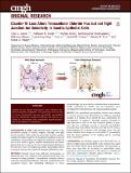
| dc.description.abstract | © 2020 The Authors Background & Aims: Tight junctions form a barrier to the paracellular passage of luminal antigens. Although most tight junction proteins reside within the apical tight junction complex, claudin-18 localizes mainly to the basolateral membrane where its contribution to paracellular ion transport is undefined. Claudin-18 loss in mice results in gastric neoplasia development and tumorigenesis that may or may not be due to tight junction dysfunction. The aim here was to investigate paracellular permeability defects in stomach mucosa from claudin-18 knockout (Cldn18-KO) mice. Methods: Stomach tissue from wild-type, heterozygous, or Cldn18-KO mice were stripped of the external muscle layer and mounted in Ussing chambers. Transepithelial resistance, dextran 4 kDa flux, and potential difference (PD) were calculated from the chambered tissues after identifying differences in tissue histopathology that were used to normalize these measurements. Marker expression for claudins and ion transporters were investigated by transcriptomic and immunostaining analysis. Results: No paracellular permeability defects were evident in stomach mucosa from Cldn18-KO mice. RNAseq identified changes in 4 claudins from Cldn18–KO mice, particularly the up-regulation of claudin-2. Although claudin-2 localized to tight junctions in cells at the base of gastric glands, its presence did not contribute overall to mucosal permeability. Stomach tissue from Cldn18–KO mice also had no PD versus a lumen-negative PD in tissues from wild-type mice. This difference resulted from changes in transcellular Cl– permeability with the down-regulation of Cl– loading and Cl– secreting anion transporters. Conclusions: Our findings suggest that Cldn18-KO has no effect on tight junction permeability in the stomach from adult mice but rather affects anion permeability. The phenotype in these mice may thus be secondary to transcellular anion transporter expression/function in the absence of claudin-18. | en_US |
| dc.language.iso | en | |
| dc.publisher | Elsevier BV | en_US |
| dc.relation.isversionof | http://dx.doi.org/10.1016/J.JCMGH.2020.10.005 | en_US |
| dc.rights | Creative Commons Attribution-NonCommercial-NoDerivs License | en_US |
| dc.rights.uri | http://creativecommons.org/licenses/by-nc-nd/4.0/ | en_US |
| dc.source | Elsevier | en_US |
| dc.title | Claudin-18 Loss Alters Transcellular Chloride Flux but not Tight Junction Ion Selectivity in Gastric Epithelial Cells | en_US |
| dc.type | Article | en_US |
| dc.contributor.department | Massachusetts Institute of Technology. Division of Comparative Medicine | |
| dc.contributor.department | Massachusetts Institute of Technology. Department of Biological Engineering | |
| dc.relation.journal | Cellular and Molecular Gastroenterology and Hepatology | en_US |
| dc.eprint.version | Final published version | en_US |
| dc.type.uri | http://purl.org/eprint/type/JournalArticle | en_US |
| eprint.status | http://purl.org/eprint/status/PeerReviewed | en_US |
| dc.date.updated | 2021-09-13T15:08:47Z | |
| dspace.orderedauthors | Caron, TJ; Scott, KE; Sinha, N; Muthupalani, S; Baqai, M; Ang, L-H; Li, Y; Turner, JR; Fox, JG; Hagen, SJ | en_US |
| dspace.date.submission | 2021-09-13T15:08:51Z | |
| mit.journal.volume | 11 | en_US |
| mit.journal.issue | 3 | en_US |
| mit.license | PUBLISHER_CC | |
| mit.metadata.status | Authority Work Needed | en_US |
