Visualization of nitric oxide production in the mouse main olfactory bulb by a cell-trappable copper(II) fluorescent probe
DownloadLippard_Visualization of nitric.pdf (362.4Kb)
PUBLISHER_POLICY
Publisher Policy
Article is made available in accordance with the publisher's policy and may be subject to US copyright law. Please refer to the publisher's site for terms of use.
Terms of use
Metadata
Show full item recordAbstract
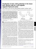
We report the visualization of NO production using fluorescence in tissue slices of the mouse main olfactory bulb. This discovery was possible through the use of a novel, cell-trappable probe for intracellular nitric oxide detection based on a symmetric scaffold with two NO-reactive sites. Ester moieties installed onto the fluorescent probe are cleaved by intracellular esterases to yield the corresponding negatively charged, cell-impermeable acids. The trappable probe Cu[subscript 2](FL2E) and the membrane-impermeable acid derivative Cu[subscript 2](FL2A) respond rapidly and selectively to NO in buffers that simulate biological conditions, and application of Cu[subscript 2](FL2E) leads to detection of endogenously produced NO in cell cultures and olfactory bulb brain slices.
Date issued
2010-05Department
Massachusetts Institute of Technology. Department of ChemistryJournal
Proceedings of the National Academy of Sciences
Publisher
National Academy of Sciences (U.S.)
Citation
McQuade, L. E., J. Ma, G. Lowe, A. Ghatpande, A. Gelperin, and S. J. Lippard. “Visualization of nitric oxide production in the mouse main olfactory bulb by a cell-trappable copper(II) fluorescent probe.” Proceedings of the National Academy of Sciences 107, no. 19 (May 11, 2010): 8525-8530.
Version: Final published version
ISSN
0027-8424
1091-6490